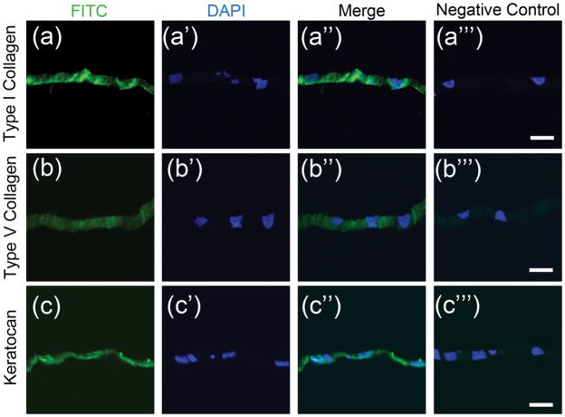
Figure 3. Immunostaining of scaffold-free tissue sheets produced in vitro.
Paraffin sections of scaffold-free tissue sheets produced after 12 days culture in KDM were immunostained as described in Methods. (a) Presence of for type I collagen (green) was detected in the matrix of scaffold-free tissue sheets, (a′) with corresponding nuclear DAPI stain (blue), (a″) merged image of (a) and (a′), and (a″′) corresponding merged image of negative control. (b) Type V collagen expression was seen in engineered tissue sheets with (b′) corresponding nuclear DAPI stain (blue), (b″) merged image of (b) and (b′), and (b″′) corresponding merged image of negative control. (c) Keratocan expression was detected in scaffold-free tissue sheets, with (c′) corresponding nuclear DAPI stain (blue), (c″) merged image of (c) and (c′), and (c″′) corresponding merged image of negative control. Scale bars: 20 μm

