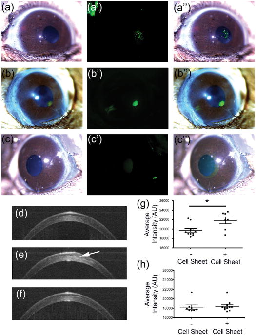
Figure 4. Histological analysis of scaffold-free tissue sheets in mouse stromal pockets in vivo.
Mouse eyes were imaged in vivo after lamellar transplantation of scaffold-free tissue sheets as described in Methods. (a) Light micrograph image of mouse eye containing scaffold-free tissue sheet with DiO-labeled cells, (a′) with corresponding fluorescent image showing human cells (green), and (a″) the merged image of (a) and (a′). (b) Light micrograph image of mouse eye containing scaffold-free tissue sheet with DTAF labelled matrix, (b′) with corresponding fluorescent image showing matrix (green) from scaffold-free sheet, and (b″) the merged image of (b) and (b′). (c) A light micrograph image of control mouse eye lacking a scaffold-free tissue sheet is shown, (c′) with corresponding fluorescent image, and (c″) the merged image of (c) and (c′). Optical coherence tomography (OCT) was used to assess light scatter in the corneal stroma. (d) Cross-sectional projection image of untreated control mouse cornea. (e) Cross sectional image of a mouse eye with implanted tissue sheet (arrow) 1 week after implantation. (f) Cross-sectional projection image of mouse eye, 5 weeks after tissue sheet implantation. (g) Quantification of light scatter from OCT scans of the stroma showing light scatter by implanted tissue sheet 1-week post-implantation. (h) Quantification of light scatter from OCT scans of the stroma showing light scatter by implanted tissue sheet 5-weeks post-implantation.

