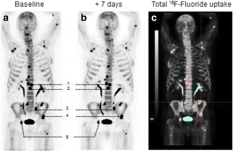
Fig. 1.

Patient 8 at baseline (a) and at +7 days (b) performing PET1 and PET2. Five chosen index lesions (numbered 1–5) were delineated for VOI measuring SUVmax, SUVmean, FTV50% and total lesion fluoride uptake at PET/CT (baseline). c Included lesions are highlighted in red (n = 17) and the blue uptake represents lesions that were omitted, urine activity and degenerative or equivocal findings
