Figure 3.

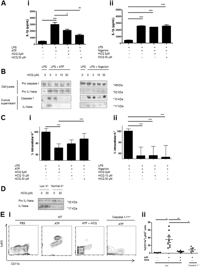
HCQ inhibits ATP-induced inflammasome activation. In vitro experiments were carried out with PMA-differentiated THP-1 macrophages. (A) Cells were primed with 0.25 µM LPS for 3 hours and then washed. When HCQ was used, it was added at this step during 15 minutes. Without further washing, cells were then treated for 45 minutes with 5 mM ATP or 2.5 µM nigericin, in the presence or not of HCQ. Culture supernatant was harvested and IL-1beta was quantified by ELISA. One experiment representative of five is shown. (B) Western blot analyses of culture supernatant of THP-1 cells treated with the indicated compounds. One experiment representative of five is shown. (C) K+ efflux was studied by quantifying intracellular K by spectroscopy under the indicated treatments. A pool of three independent experiments is shown. (D) Western blots studying IL-1beta maturation in cells cultured during 45 minutes with physiological buffer or without K+. One experiment representative of two is shown. (E) WT or caspase-1−/− C57Bl/6 mice were injected with 20 mg/kg ATP. 4 hs later, peritoneal lavage was performed and cells were stained with anti-CD11b and Ly6G antibodies. In HCQ-treated animals, 1 mg/kg of the drug was injected i/p daily 7 days before ATP injection. i) Representative dot plots are shown. ii) Neutrophil quantification from two independent experiments. *p < 0.05; **p < 0.01; ***p < 0.001.
