Figure 2.
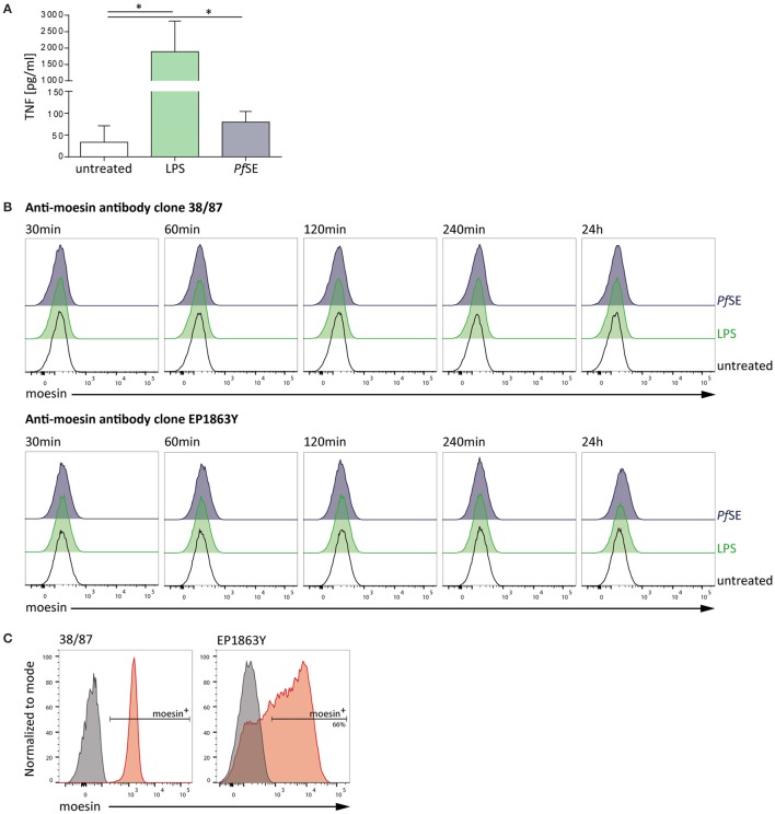
Macrophage-like THP-1 cell response to stimulation is not accompanied by moesin cell surface localization. (A) Macrophage-like THP-1 cell responsiveness to stimulation determined by TNF-ELISA after 24 h treatment; LPS: 10 ng/ml, P. falciparum schizont extract (PfSE): 1:100 in medium; n = 5, bars indicate mean + SD; One-way ANOVA of paired samples with Dunnett correction revealed *P < 0.05. (B) Representative histograms of moesin cell surface staining in macrophage-like THP-1 cells at different time points of stimulation, detected by flow cytometry with time points and treatments indicated, gated on viable CD11b+ cells, detection with anti-moesin antibody clone 38/87, and anti-mouse IgG-FITC [upper panel; cells differentiated with 20 ng/ml PMA for 24 h (Iontcheva et al., 2004))] and with anti-moesin antibody clone EP1863Y and anti-rabbit IgG-PE (lower panel; cells differentiated with 200 nM PMA for 72 h) (C) Intracellular staining control for moesin; left: viable undifferentiated THP-1, right: viable differentiated THP-1; gray: secondary antibody staining control, red: moesin+ cells detected with anti-mouse IgG-APC for clone 38/87, and anti-rabbit IgG-PE for clone EP1863Y. Dead cells were excluded from all flow cytometric analyses by using a fixable viability dye.

