Figure 4.
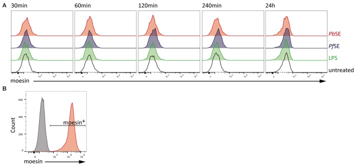
Absence of moesin on BMDM cell surface. (A) Flow cytometric analysis of moesin cell surface localization on viable CD11b+F4/80+ BMDM over time with stimuli indicated; detection of anti-moesin EP1863Y using anti-rabbit-PE; (B) Intracellular moesin detection in viable CD11b+ BMDM; gray: secondary antibody control, red: moesin+ cells using anti-moesin antibody EP1863Y and anti-rabbit IgG-AlexaFluor488.

