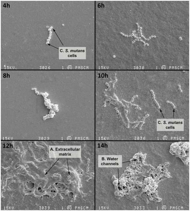
FIGURE 3.
Scanning electron microscopy (SEM) of the single-species biofilm formed by S. mutans cells at various time points. 4–6 h – initial phase of adhesion of S. mutans to a flat surface of polystyrene; S. mutans adheres to the polystyrene surface primarily using a sucrose-dependent mechanism (based on the glucosyltransferase and glucan-binding proteins); 8–14 h – biofilm occurrence, at this stage there is an irreversible merger of the bacteria with the surface and then the formation of the extracellular matrix (ECM) which protects against host defense factors and drying; 16 h – biofilm maturation, during which the matrix is still being formed and other bacterial species also attach to the biofilm, bacteria synthesize extracellular polymers (soluble and insoluble glucans, fructans, and heteropolymers) which are components of the plaque matrix. The appearance of the matrix is a feature of all biofilms, but it is more than a chemical scaffold that maintains the biofilm shape. The matrix is biologically active and retains water, nutrients and enzymes within the biofilm structure: A – polymeric ECM, which has an open architecture with nutrient channels, spaces, and other properties, e.g., environmental heterogeneity (pH and oxygen gradients, co-adhesion); B – water channels; C – S. mutans cell. The original magnification: 4000x.

