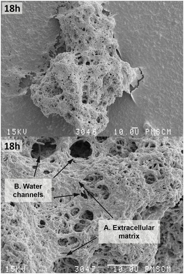
FIGURE 4.
Scanning electron microscopy of the single-species mature biofilm formed by S. mutans cells after 18 h. The photo shows S. mutans cells (C) forming a mature biofilm with a visible ECM (A) constituting the scaffold of the entire biofilm structure with nutrient channels, (B) and other biofilm properties like water channels. The original magnification: 400x, 2000x.

