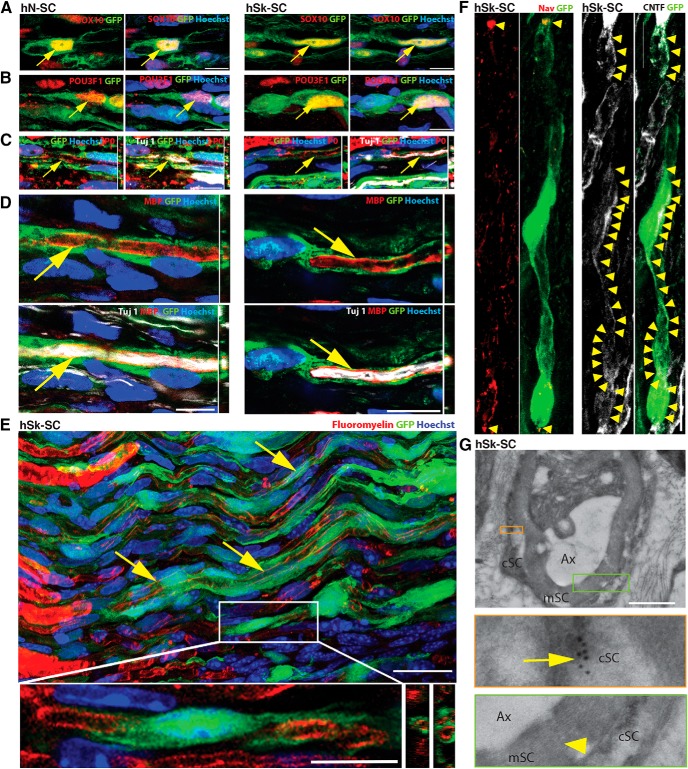
Figure 6.
Adult human nerve and skin-derived SCs are able to ensheath rodent axons and produce limited myelin. SCs were transplanted into nerve injury and assessed by immunofluorescence at five to eight weeks after injury. A–F, Representative images show GFP-positive adult human nerve (hN-SC) and skin (hSk-SC) SCs (green) coexpressing (red, arrows) the pan-SC protein, SOX10 (A), the promyelinating factor, POU3F1 (B), the myelin proteins, P-zero (C) and MBP (D), and the myelin lipid marker, Fluoromyelin (E). Insets show higher magnification image of SCs and Z-plane view of the close association of myelin proteins with GFP. We also noted the presence of Nav1.6+ sodium channels at the nodes (red, arrowheads; F), and CNTF+ mesaxons throughout the internode (white, arrowheads; F), as well as GFP+ immunogold particles (inset, arrow; G) associated with compact myelin (inset, arrowhead; G). Scale bar: 10 μm (A–F), 25 μm (E), 1 μm (G); Ax, axon; cSC, SC cytoplasm; mSC, myelin (G). Representative images are from 46- and 27-year-old male.

