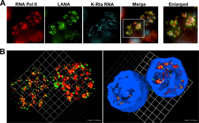
FIG 6.
(A) RNA-FISH showing the assembly of LANA dots with RNA Pol II in BCBL-1. RNA Pol II (red immunofluorescence), LANA (green immunofluorescence), and K-Rta RNA (light blue, RNA-FISH) were visualized. BCBL-1 was stimulated with TPA and sodium butyrate for 4 h. Cells were fixed after 24 h after the end of stimulation. Merge and enlarged merge images overlaid with K-Rta RNA staining are shown. (B) 3D view of KSHV transcriptional factory. Z-stack images were taken, and 3D images without or with DAPI staining were constructed. Green, LANA; red, RNA Pol II; orange, K-Rta transcripts; blue, DAPI.

