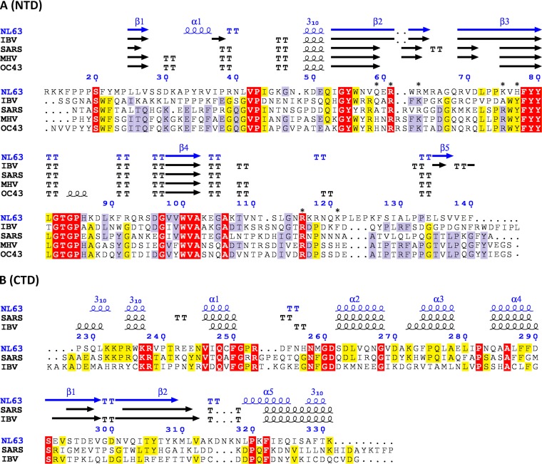
FIG 1.
Structure-guided alignment of the NTD (A) and CTD (B) of N protein. Secondary structures are indicated above the alignment. The regions of highest sequence conservation are highlighted. The residues which involvement in nucleic acid binding was tested by mutagenesis in this study are marked with asterisks. TT, turn.

