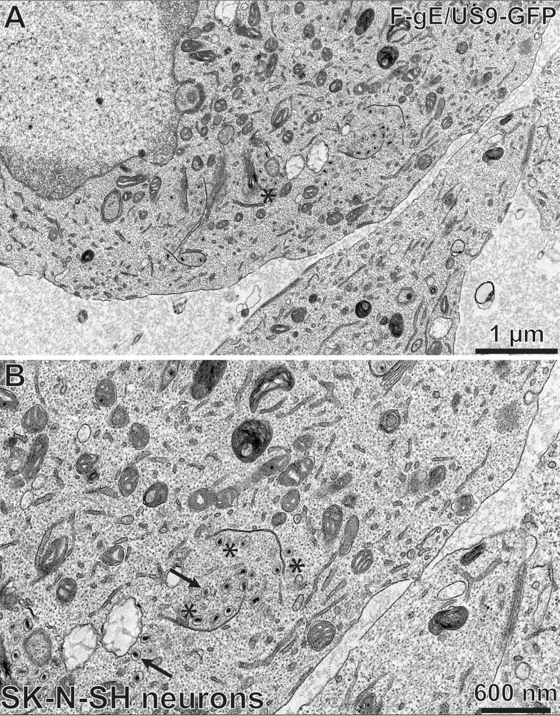
FIG 11.
Electron micrographs of SK-N-SH neurons infected with the HSV F-gE/US9-GFP double mutant. SK-N-SH neurons growing on collagen-coated 60-mm dishes were differentiated for 10 days and then infected with the gE/US9 double mutant virus F-gE/US9-GFP at 5 PFU/cell for 18 h. The cells were fixed and processed for electron microscopy. These cells contained few enveloped virions on cell surfaces (A) and numerous unenveloped or only partially enveloped capsids in the cytoplasm (B). The black arrows indicate unenveloped capsids, and asterisks indicate capsids abutting tubular membranes. In panel A, there are capsids adjacent to a stack of membranes which is likely the Golgi apparatus.

