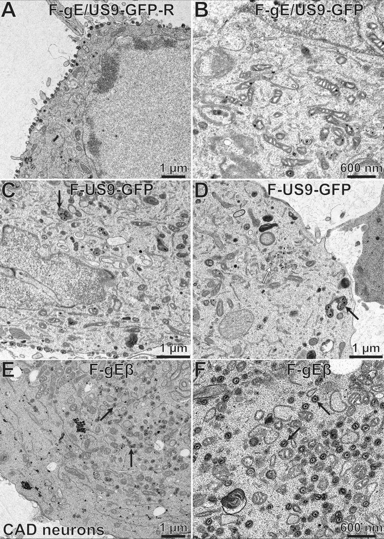
FIG 12.
Electron micrographs of CAD neurons infected with F-gE/US9-GFP-R, F-gE/US9-GFP, F-US9-GFP, and F-gEβ. CAD neurons growing on collagen-coated 60-mm dishes were differentiated for 7 days and then infected with the repaired virus F-gE/US9-GFP-R (A), the double mutant F-gE/US9-GFP (B), a mutant lacking just US9 (F-US9-GFP) (C and D), or a mutant lacking just gE (F-gEβ) (E and F) at 20 PFU/cell. After 18 h, the cells were fixed and processed for electron microscopy. CAD neurons infected with F-gE/US9-GFP-R exhibited largely enveloped virions at the cell surface, while cells infected with F-gE/US9-GFP showed numerous unenveloped (white arrow in panel B) or partially enveloped capsids in the cytoplasm. CAD neurons infected with the US9 mutant F-US9-GFP exhibited numerous dense vesicles, frequently containing several capsids (black arrows in panels C and D), as well as unenveloped capsids (white arrow in panel D). CAD cells infected with the gE mutant F-gEβ exhibited primarily fully enveloped capsids in the cytoplasm (black arrows in panels E and F), with few virions on the cell surface.

