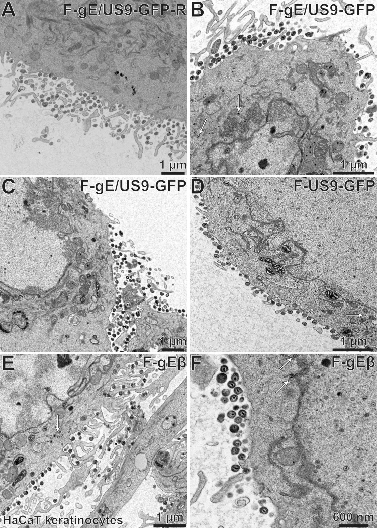
FIG 13.
Electron micrographs of HaCaT cells infected with F-gE/US9-GFP-R, F-gE/US9-GFP, F-US9-GFP, or F-gEβ. HaCaT keratinocytes were grown on plastic 60-mm dishes and then infected with the repaired virus F-gE/US9-GFP-R (A), the double mutant F-gE/US9-GFP (B and C), the US9 mutant US9-GFP (D), or the gE mutant F-gEβ (E and F) at 15 PFU/cell. After 18 h, the cells were fixed and processed for electron microscopy. All four of these viruses produced largely cell surface virions, though there was some accumulation of unenveloped capsids (white arrows) with the F-gE/US9-GFP double mutant and the gE-null mutant (F-gEβ). No significant defects in assembly were observed with the US9 mutant (F-US9-GFP).

