FIG 2.
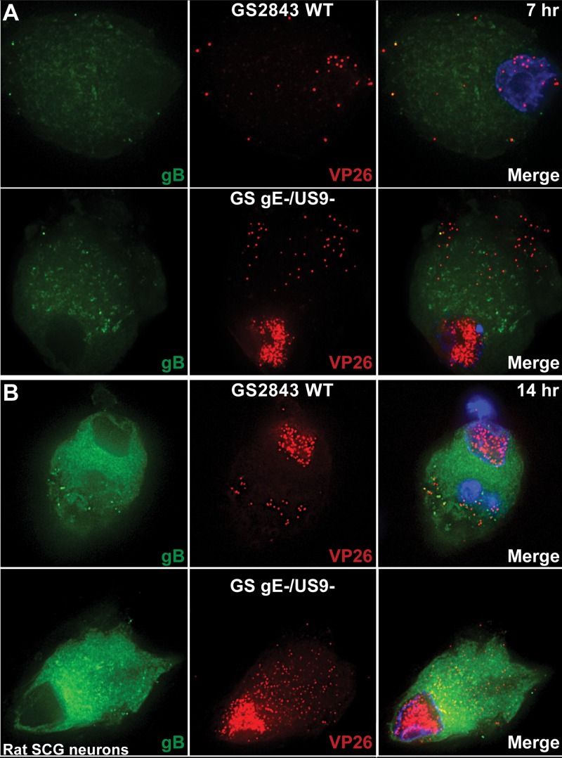
Immunofluorescence imaging of HSV capsids and gB in rat embryonic SCG neurons. Rat SCG neurons grown on polylysine/laminin-coated glass coverslips were infected with wild-type (WT) HSV GS2843 expressing gB-GFP and the capsid protein VP26-RFP or with a derivative of this virus lacking both the gE and US9 genes (GS gE− US9−) at 8 PFU/cell. Cells were fixed with paraformaldehyde after 7 or 14 h and then permeabilized, and nuclei were stained with 300 nM DAPI. At 7 h (A) and 14 h (B), substantially more cytoplasmic capsids were observed with GS gE− US9− than with wild-type GS2843.

