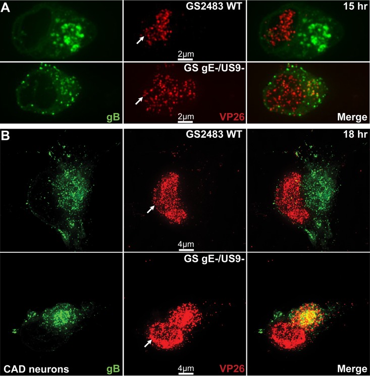
FIG 4.
Immunofluorescence imaging of capsids and gB in CAD neurons infected with wild-type HSV GS2843 or with GS gE− US9−. CAD neurons growing on polylysine/laminin-coated glass coverslips were differentiated for 8 days, infected with wild-type GS2843 expressing gB-GFP and VP26-RFP or with GS gE− US9− at 8 PFU/cell, and fixed with 4% paraformaldehyde at 15 h and 18 h postinfection. At 15 h (A) and 18 h (B), more cytoplasmic capsids accumulated in the cytoplasm of CAD cells infected with the GS gE− US9− double mutant than in that of WT GS2843-infected cells. White arrows in the VP26 column indicate the position of the nucleus. Note that the yellow color in the merged panels is not representative of colocalization but resulted from the flattened z-stacks used to produce the image.

