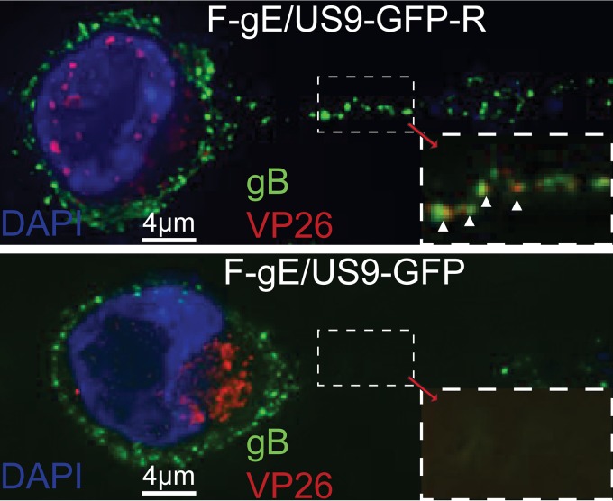
FIG 5.
Immunofluorescence imaging of CAD neuron axons. CAD neurons growing on polylysine/laminin-coated glass coverslips were differentiated for 9 days and then infected with HSV F-gE/US9-GFP-R, in which the gE and US9 genes were repaired, or with the F-gE/US9-GFP double mutant lacking gE and US9, at 8 PFU/cell. At 18 h postinfection, the cells were fixed with 4% paraformaldehyde, permeabilized, immunostained with antibodies specific for HSV gB (green) and VP26 (red), and also stained with 300 nM DAPI (blue). CAD cells infected with F-gE/US9-GFP exhibited many capsids in the cytoplasm and very few or no capsids in the axon, whereas neurons infected with F-gE/US9-GFP-R showed few cytoplasmic capsids and capsids in axons. White arrowheads point to enveloped (married) virions in axons.

