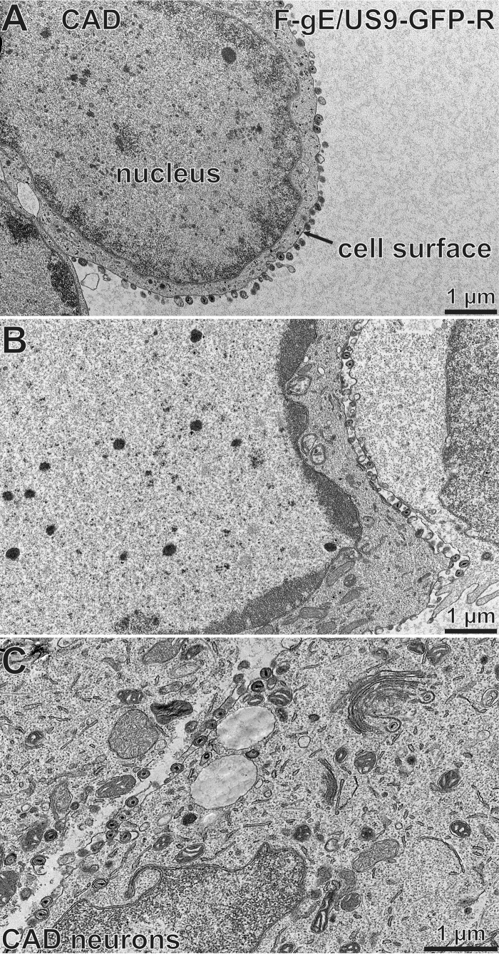
FIG 6.
Electron micrographs of CAD neurons infected with the repaired HSV strain F-gE/US9-GFP-R. CAD neurons growing on collagen-coated 60-mm dishes were differentiated for 7 days and then infected with the repaired virus, F-gE/US9-GFP-R, at 20 PFU/cell for 18 h. The cells were then fixed and processed for electron microscopy. The majority of the capsids were enveloped and on cell surfaces. The cell surface and nucleus are indicated in panel A.

