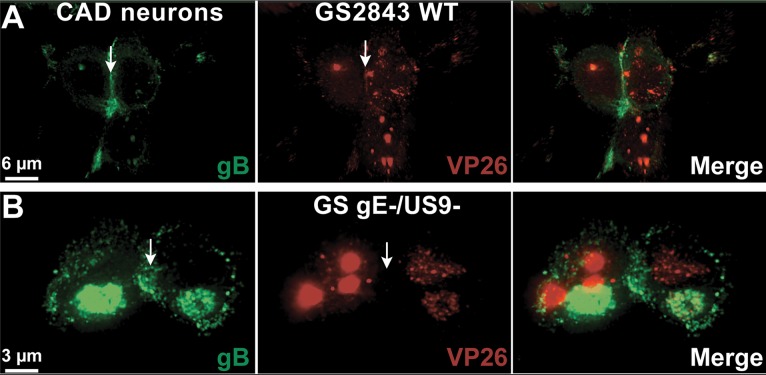
FIG 8.
Immunofluorescence imaging of CAD neurons infected with GS2843 or GS gE− US9−, focusing on cell-cell junctions. CAD neurons growing on polylysine/laminin-coated glass coverslips were differentiated for 10 days and then infected with WT GS2843 expressing gB-GFP and VP26-RFP (A) or with the gE− US9− version of this virus (GS gE−/US9−) (B) for 18 h. The cells were then fixed with 4% paraformaldehyde and analyzed by confocal microscopy. For these images, 16 separate 0.2-μm confocal sections were stacked together into a flattened image to attain images of cell-cell junctions. Numerous capsids accumulated at cell-cell junctions (white arrows) in cells infected with WT GS2843, but this was not the case for cells infected with the GS gE− US9− double mutant.

