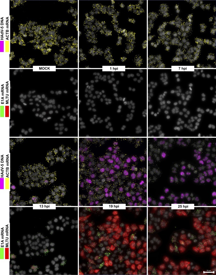
FIG 3.
Temporal expression pattern of HAdV-5 mRNAs in infected HeLa cells. HeLa cells infected with HAdV-5 (10 FFU/cell) and analyzed during different time points (1, 7, 13, 19, and 25 hpi). HAdV-5 genomic DNA (magenta) is presented along with ACTB mRNA (yellow). E1A mRNAs (13S and 12S, green) are presented along with MLTU mRNAs (exon I_II and exon II_III, red). Scale bar, 50 μm.

