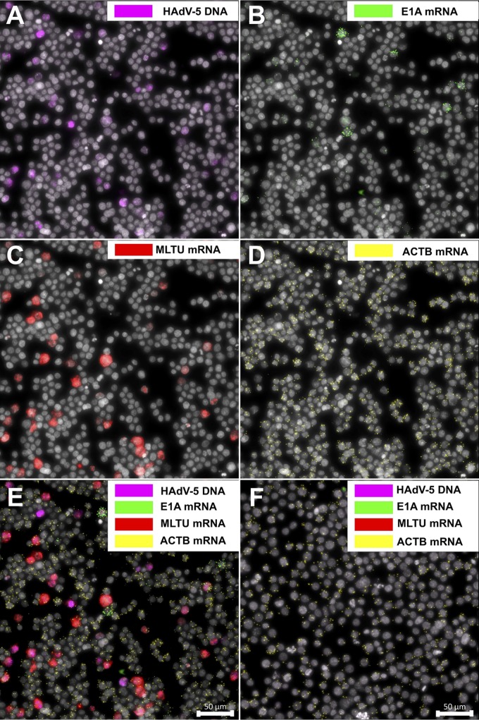
FIG 6.
Concurrent detection of HAdV-5 DNA and mRNAs in long-term infected BJAB cells. BJAB cells were infected with HAdV-5 (100 FFU/cell) and analyzed 6 days postinfection (dpi). HAdV-5 DNA is presented in magenta (A), E1A mRNAs in green (B), MLTU mRNAs in red (C), and ACTB mRNA in yellow (D). (E) Merged image. (F) Uninfected cells stained with the aforementioned padlock probes. Scale bar, 50 μm.

