FIG 8.
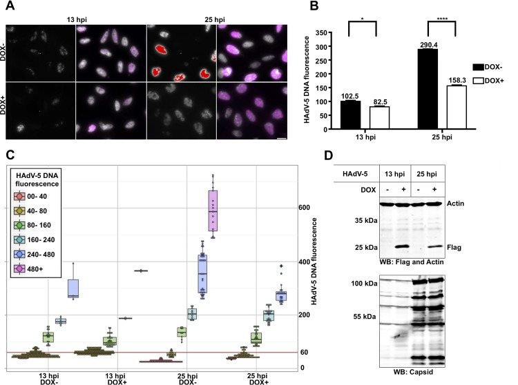
Expression of the pVII(Δ24) protein reduces HAdV-5 DNA detection with RCA. (A) HeLa-pVII(Δ24)Flag cells were treated with doxycycline (Dox+) or were left untreated (Dox−) for 24 h, followed by HAdV-5 infection in the presence or absence of Dox. RCA with PLP recognizing HAdV-5 genomic DNA (magenta or raw black-and-white exposure) at 13 hpi and 25 hpi is shown. The scale bar is 20 μm. (B) Geometric averages and standard deviations of HAdV-5 DNA signal. Average HAdV-5 DNA fluorescence was plotted against infection time (x axis). ****, P ≤ 0.0005; *, P ≤ 0.05. (C) Single-cell HAdV-5 DNA fluorescence in HeLa cells 13 hpi and 25 hpi. Cells were divided into groups and color coded based on their HAdV-5 DNA fluorescence. Each data point (which represents an individual cell) was distributed in the plot depending on the time point, Dox treatment (x axis), and HAdV-5 DNA fluorescence (y axis). A box-and-whisker plot for each cell group was overlaid with data points. The red line indicates an HAdV-5 DNA fluorescence of 60, showing the fluorescence cutoff used to exclude uninfected cells to generate the accurate and unbiased averages and standard deviations shown in panel B. (D) Detection of the pVII(Δ24)Flag protein with anti-Flag antibody by Western blotting (WB).

