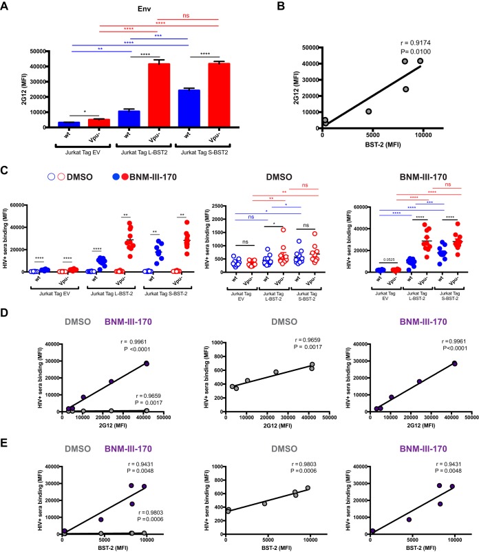
FIG 2.
BST-2 expression correlates with cell surface Env level and recognition of HIV-1-infected cells by HIV+ sera. Jurkat Tag cells expressing no BST-2 (Jurkat Tag EV [empty vector]) or stably expressing L-BST-2 or S-BST-2 were mock infected or infected with the transmitted/founder virus HIV-1 CH58 (CH58 T/F) expressing Vpu (wild-type CH58 T/F [wt]) or not expressing Vpu (Vpu−). Forty-eight hours postinfection, cells were stained with the anti-Env Ab 2G12 (A and B) or with sera from 10 HIV-1-infected individuals (C) in the presence of the CD4mc BNM-III-170 (50 μM) or equivalent volume of DMSO, followed with appropriate secondary Abs. (A) Mean fluorescence intensity (MFI) of 2G12 binding obtained in at least six independent experiments; (B) correlation between 2G12 binding and BST-2 level. (C) MFIs obtained with all the different sera in the presence of CD4mc BNM-III-170 or DMSO; (D and E) correlation between HIV+ serum binding and 2G12 level or BST-2 level, respectively, in the presence of 50 μM CD4mc BNM-III-17 or DMSO. Values are means plus standard error of the means (SEM) (error bars). Statistical significance was tested using an unpaired t test (A), a Pearson correlation test (B, D, and E), or a paired t test or Wilcoxon matched-pair signed-rank test based on statistical normality (C) (*, P < 0.05; **, P < 0.01, ***, P < 0.001; ****, P < 0.0001; ns, nonsignificant).

