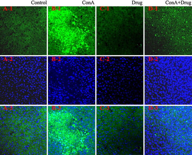
Fig. 4.

Analysis of liver cell apoptosis. Mice were sacrificed at 24 h after Con A injection. The livers were harvested from control (A), Con A (B), drug (C) and ConA+ drug (D) groups respectively. Liver tissues from the four groups were fixed and stained with TUNEL (1), DAPI (2) and overlap (3). Original magnification ×200
