Fig. 2.

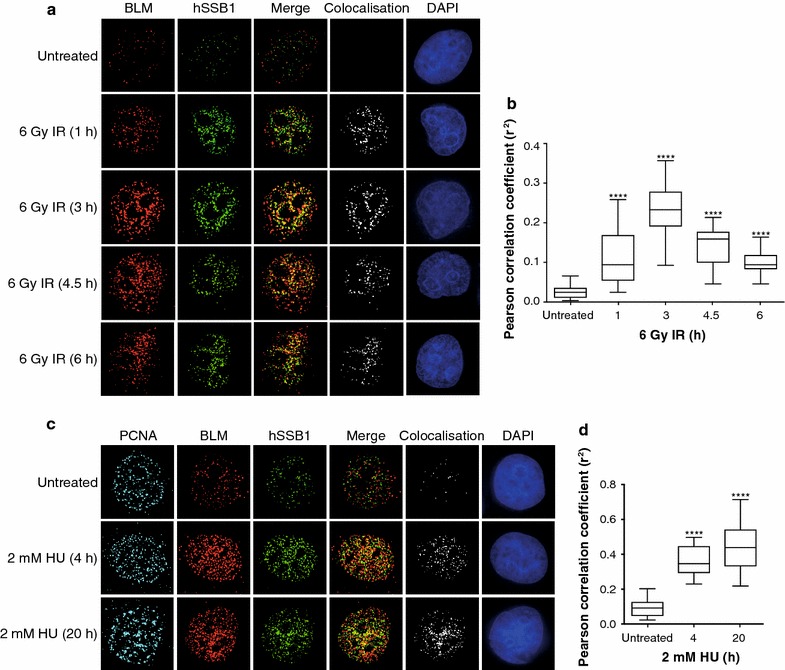
hSSB1 co-localises with BLM at IR- and HU-induced nuclear foci. a, c HeLa cells were pre-extracted prior to fixation, 0, 1, 3, 4.5 and 6 h post ionising radiation (IR) treatment (a) or 0, 4 and 20 h post treatment with hydroxyurea (HU) as indicated (c). Fixed cells were incubated with primary antibodies against BLM, hSSB1 and PCNA (c only), which were detected with fluorescent secondary antibodies and visualised using a DeltaVision PDV microscope. Co-localisation of BLM and hSSB1 is demonstrated by the merged image (merge) and by the analysis of images using the Image J colocalisation plugin showing only the colocalised pixels (colocalisation). DAPI was used to stain the nuclei. b, d Box and Whisker plots representing Pearson correlation coefficient (r2) from 24 (for a) and 15 (for c) individual cells for each time-point from a and c. One-way Anova followed by post hoc analysis was used to assess the statistical significance between the mean r2 values of each treatment group and untreated cells. ****p < 0.0001
