Fig. 1.

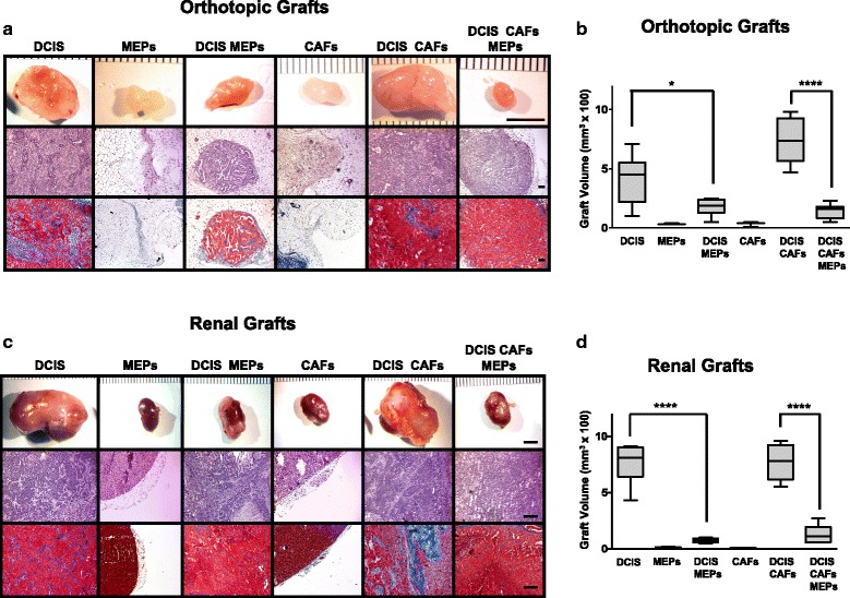
Myoepithelial cells (MEPs) reduce volume and malignant phenotype of ductal carcinoma in situ (DCIS) xenografts. MCF10.DCIS (DCIS) cells, N1ME cells (MEPs), and/or WS-12T (cancer-associated fibroblasts [CAFs]) were implanted under the renal capsule or orthotopically within the mammary fat pad of female severe combined immunodeficiency mice and evaluated after 8 weeks. a Representative orthotopic xenografts. Scale bar = 5 mm (top row); hematoxylin and eosin (H&E) staining; scale bar = 100 μm (middle row); and Masson’s trichrome staining for collagen (blue); scale bar = 100 μm (bottom row). b Volume of orthotopic xenografts (n = 3–8). c Representative renal xenografts. Scale bar = 5 mm (top row); H&E staining; scale bar = 100 μm (middle row); and Masson’s trichrome staining for collagen (blue); scale bar = 100 μm (bottom row). d Volume of renal xenografts (n = 6, except for CAFs alone, which were n = 2 and not used in statistical analyses). Data are presented as box-and-whisker plots where the box represents the interquartile range and whiskers represent minimum and maximum values. * p ≤ 0.05; **** p ≤ 0.0001 as determined by one-way analysis of variance and Bonferroni’s multiple comparisons test. Immunostaining and immunoblotting results for basal markers are shown in Additional file 1: Figure S1
