
Figure 2.
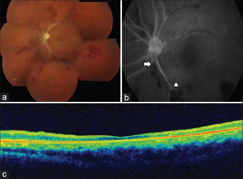
Combined occlusion of central retinal artery and vein. (a) Fundus image (montage) shows pale disc, sclerosed vessels, and intraretinal hemorrhages in all quadrants suggestive of combined occlusion, (b) fundus fluorescein angiography shows occluded artery (arrow head) and occluded vein (arrow), (c) optical coherence tomography shows thin and atrophic retinal layers suggestive of a long-standing vascular occlusion