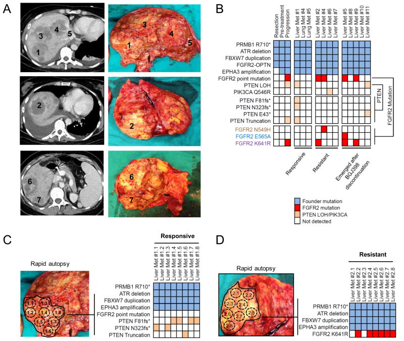
Figure 3. Rapid autopsy reveals inter-lesional and intra-lesional heterogeneity of resistance.
A. Axial contrast-enhanced CT images and autopsy photographs of seven liver metastases from Patient #'2. B. Heatmap illustrating mutations detected in the indicated autopsy lesions. Three distinct FGFR2 point mutations and four distinct PTEN mutations were identified. C and D. Corresponding images of Liver Met #1 (C) and Liver Met #2 (D) taken from Patient #2's autopsy including heatmaps indicating mutations identified in eight spatially distinct pieces isolated from Responsive Liver Met#1 and eight spatially distinct pieces isolated from Resistant Liver Met #2.

