. 2017 Feb 16;17(6):1137–1148. doi: 10.1039/c6lc01450j

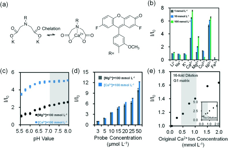
Fig. 4. Quantification of divalent metal ions in buffer solution (Tris, pH 7.4, 150 mmol L–1) at 24 °C. (a) Chelation mechanism of o-acetanisidide with divalent metal ions. (b) Fluorescence readouts of o-acetanisidide (λ ex/λ em: 485/528 nm) in the presence mono/divalent metal ions in aqueous solutions (n = 3). (c) The effect of pH on fluorescence readouts of o-acetanisidide (25 μmol L–1) at constant Mg2+ and Ca2+ ion concentrations (100 mmol L–1) in aqueous solutions (n = 3). (d) Fluorescence intensity readouts of Mg2+ and Ca2+ ions (100 mmol L–1) as the concentration of o-acetanisidide were varied from 3–50 μmol L–1 (n = 3). (e) Quantification of 16-fold diluted Ca2+ ions on G1 matrix at a constant o-acetanisidide concentration (25 μmol L–1) (n = 6). Insets show the quantification of non-diluted Ca2+ ions on G1 matrix (n = 3). Shadows in Fig. 4c and e show the physiological pH and Ca2+ ion concentration range. Error bars represent standard error of the mean.

Fig. 4