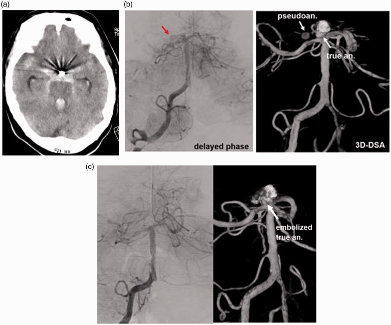
Figure 4.
(a) CT showing SAH. (b) Angiography demonstrating a cavity protruding to the right from the recurrent BA tip aneurysm. Angiography showing delayed filling and washout of CM in the additional cavity. (c) Post-embolization angiography demonstrating complete obliteration of the aneurysm and disappearance of the pseudoaneurysm.
CT: computed tomography; SAH: subarachnoid hemorrhage; CM: contrast medium.

