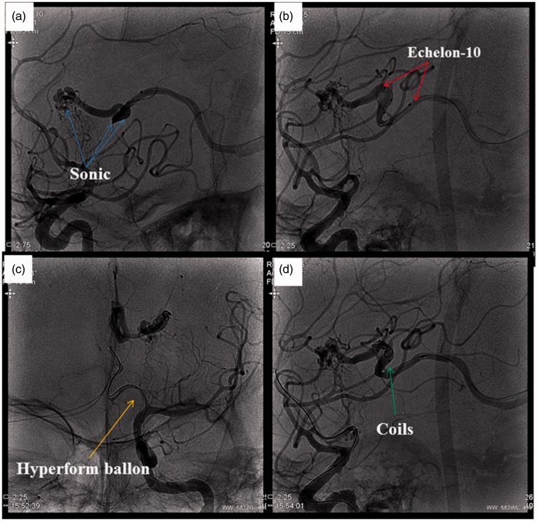
Figure 4.
(a) A microcatheter (Sonic, Balt, France) was placed as close as possible to the nidus. (b) Another microcatheter (Echelon-10, Ev3, Irvine, California, USA) was navigated alongside the Sonic into the draining vein. Its tip was positioned between the most distal marker and the detachment zone of the Sonic. (c) A balloon was placed from the anterior cerebral artery to the end of the carotid artery. (d) Then, coils were placed in the draining vein through the Echelon-10 microcatheter to create a plug.

