
Figure 2. Immunolocalization of MIP-2 and CXCR2 in 13.5 day embryos.
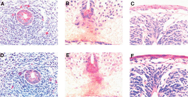
The esophagus stained positive for MIP-2 (A) and CXCR2 (D). The floor plate of the neural tube was positive for MIP-2 (B) and CXCR2 (E). C and F show the immunostaining for MIP-2 (C) and CXCR2 (F) in the ectoderm of the back skin.