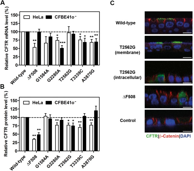
Fig 1. Synonymous mutations alter cystic fibrosis transmembrane conductance regulator (CFTR) expression.
(A) Quantitative real-time PCR (qRT-PCR) quantification of steady-state CFTR mRNA expression in HeLa and CFBE41o- cells analyzed 24 h after transfection and normalized to neomycin phosphotransferase (NPT). The mRNA level of wild-type CFTR was set to 100%. (B) Total steady-state CFTR expression (i.e., the sum of bands B and C in S1B Fig) normalized to that of NPT and β-actin (ACTB) and related to wild-type CFTR, which was set to 100%. In A and B, data are means ± SEM (A: HeLa, n = 3–7; CFBE41o-, n = 4–9; B: HeLa, n = 5–14; CFBE41o-, n = 4–7); * P < 0.05, ** P < 0.01, *** P < 0.001 versus wild-type CFTR. (C) Representative immunostaining images (n = 3) of well-differentiated human cystic fibrosis (CF) airway epithelia. The frequency of intracellular versus membrane-localized T2562G-CFTR staining was as follows: 44% of T2562G-CFTR–expressing cells exhibited intracellular staining (12 out of 27) and in 55% (15 out of 27), staining was membrane localized. The perinuclear staining demonstrates the intracellular localization of T2562G-CFTR. The control was non–CFTR-expressing epithelia. β-catenin immunostaining denotes adherens junctions and DAPI—nuclei; scale bars, 10 μm. The underlying data of panels A and B can be found in S1 Data.

