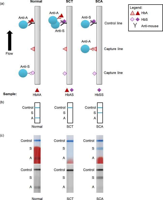
Fig 1. Schematic and example images of lateral flow strips.
(a) Schematic of lateral flow test strip for three possible conditions: normal, sickle cell trait, and sickle cell disease blood. HbA, HbS, and an anti-mouse control antibody are dried on the strip at the capture and control lines. Two populations of latex beads (one conjugated to anti-HbA, the other to anti-HbS) and the blood sample are flowed up the strip. (b) Resulting visible readout on the strip for normal, sickle cell trait, and sickle cell disease blood. (c) Scanned images of example strips run with patient blood. Top image shows full-color scan; bottom image shows red channel of same image. Labels on the side are a guide to interpreting the competitive assay: a line present at the “A” line indicates normal, at the “S” line indicates SCA, and at neither indicates SCT. The positive control line must be present for the test to be valid.

