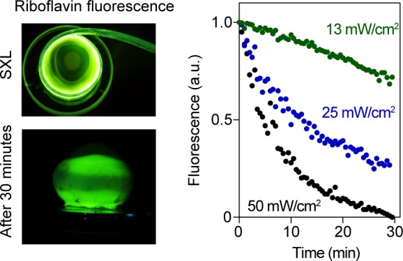
Figure 5.

Photobleaching of riboflavin-stained sclera. Left: Images of porcine eyeballs during (top) and after (bottom) SXL with blue light. Bottom image shows pattern of bleached riboflavin along the equator after 30 minutes of irradiation. Right: Photobleaching of riboflavin, using mean irradiances of 13 to 50 mW/cm2.
