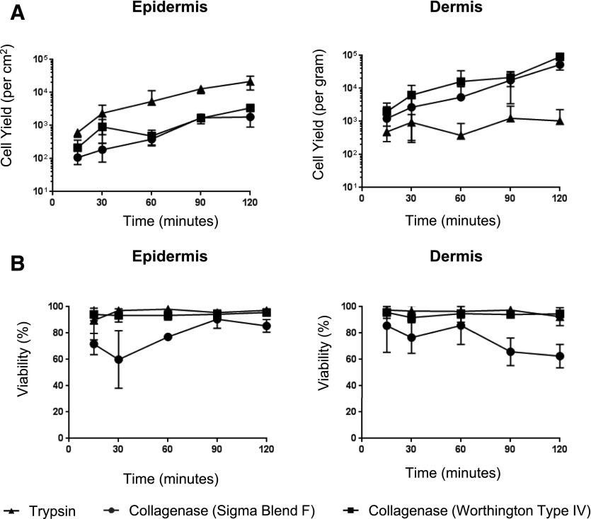
Figure 2. Identification of the optimal enzyme for the isolation of mononuclear phagocytes from skin.
Epidermal and dermal tissue was processed as shown in Fig. 1. Each tissue sample was split into roughly 4 equal parts and weighed before digestion with 0.5% trypsin or 200 U collagenase (Worthington type II or IV or Sigma blend F) at 37°C; 1 ml of cell suspension was harvested at 10–30-min intervals for 120 min, and true count beads were added before staining with Live/Dead NIR, HLA-DR PerCP, and CD45 PE.Cy7, and cell suspension was analyzed by flow cytometry for cell yield (A) and viability (B). n = 3. The data obtained using type II collagenase was similar to the data for blend F. All enzymatic digestion comparisons were performed on tissue from the same donor.

