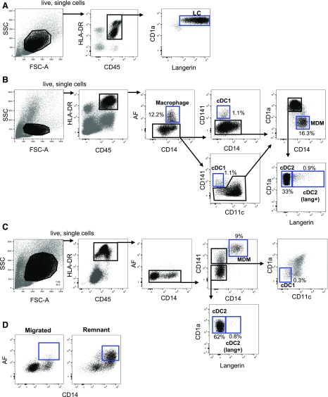
Figure 3. Identification of mononuclear phagocytes from human skin by multiparameter flow cytometry.
Cells were isolated from skin with either collagenase digestion (A and B) or spontaneous migration (C). Cells were stained with Live/Dead NIR, HLA-DR BUV395, CD45 BV786, CD1a BV510, CD3 AF700, CD19 BV605, CD11c PE.CF594, CD14 BUV737, CD141 BV711, and langerin vioblue. All epidermal and dermal cells were gated within the live, single HLA-DR+CD45+CD3−CD19− population. (A) Langerhans cells were defined as the CD1a+ langerin+ cells in the epidermis. (B) Dermal mononuclear phagocytes were gated in sequential order. Macrophages were defined as the autofluorescent CD14+ cells. Within the nonautofluorescent population, cDC1s were defined as the CD141+CD14− or CD141+CD11clow population. The CD141− population was split into CD14+ CD1a− cells, MDMs, and 2 populations of CD1a+CD14− cDC2s, that could be distinguished by langerin expression. (C) Three populations of nonautofluorescent, dermal, migratory cells could be distinguished; CD14+ MDMs were defined as CD141high CD14+. The CD141low population was gated on the 2 populations of cDC2s. The CD141+CD14− population was gated, and cDC1s were then defined as CD11c−CD1a− cells within that gate. (D) CD14+ autofluorescent macrophages were present in negligible amounts in the migrated populations but were able to be liberated from tissue by collagenase digestion after 48 h of culture. Representative flow cytometry data are shown, and the relative proportion of each cell subset from the HLA-DR+CD45+ gate is shown. (A and B) n = 50, (C) n = 15, (D) n = 4.

