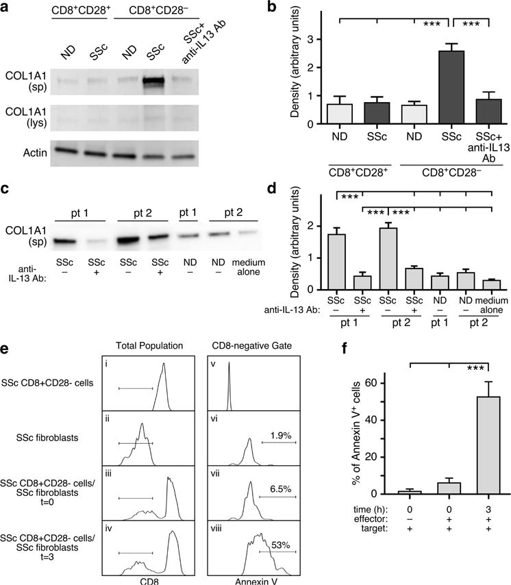
Figure 5. SSc CD8+CD28− T cells are pro-fibrotic and cytotoxic to dermal fibroblasts in vitro.

(a–b) Normal dermal fibroblasts were stimulated for 24 hours with CD8+CD28− and CD8+CD28− T-cell subset supernatants from ND (n=7) or SSc patients (n=8) with or without pre-incubation with a neutralizing anti-IL-13 antibody. COL1A1 production was determined in culture supernatants (sp) or cell lysates (lys) by Western blot. A representative experiment out of seven independent experiments from different SSc patients is shown. SSc dermal fibroblasts were obtained from two early dcSSc patients (pt 1 and 2) and were cultured with ND (n=8) and SSc (n=10) CD8+CD28− T-cell supernatants. (c) Representative Western blot for COL1A1 protein expression in fibroblast culture media (sp) and densitometric quantification of secreted COL1A1 protein (d). SSc CD8+CD28− cells (ei) or fibroblasts (eii) alone as well as mixed at a 5:1 ratio (eiii, eiv) were incubated in media for 0 or 3 hours. Lymphocyte and fibroblast populations were gated according to light scatter characteristics (FSC/SSC). The horizontal lines in panels ei–eiv designate the CD8-negative target cell gate used for analysis of Annexin V positivity in panels ev–eviii. This experiment is representative of three similar experiments. Percent Annexin V-positive cells was determined by population gating (f). Statistics in b,d, and f by ANOVA followed by post hoc Tukey’s test.
