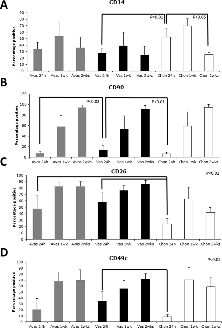
Figure 1.
Surface molecule profiles (percentage positive) over time in culture on cells derived from either human avascular (Avas), vascular (Vas) meniscus tissue, or from human articular cartilage (Chon). A. CD14 levels did not significantly change on meniscus cells. A significant reduction on chondrocytes noted by 2 weeks in monolayer culture. B. Vascular-derived meniscus cells possessed significantly higher levels of CD90/THY-1 compared to the avascular-derived meniscus cells (24 h). C. A significantly higher percentage of CD26 positive cells from meniscus was detected in early culture (24 h). D. Significantly higher percentage alpha 3 integrin (CD49c) was detected on cells isolated from the vascular region of meniscus compared to chondrocytes at 24 h.

