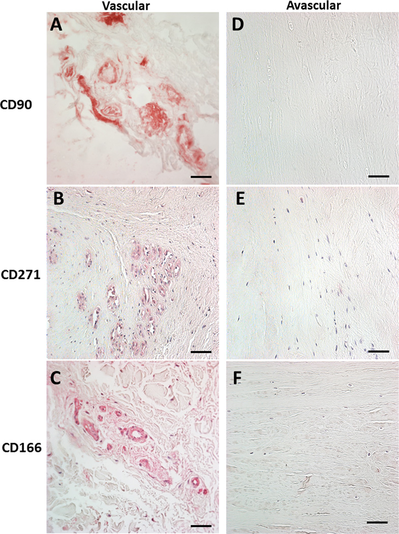
Figure 3.
Immunohistochemistry of the vascular meniscus tissue region for multiple progenitor markers. Positive signals are associated with blood vessels indicating the presence of pericytes. No positive signals were seen in the avascular regions, with the exception of the surface of the meniscus spanning all regions (see Figure 4). A. CD90 / THY-1 in the vascular region. B. CD271 / LNGFR in the vascular region. C. CD166 /ALCAM in the vascular region. D. CD90 / THY-1 in the avascular region. E. CD271 / LNGFR in the avascular region. F. CD166 /ALCAM in the avascular region. All images 40x magnification (scale bar 50 µm).

