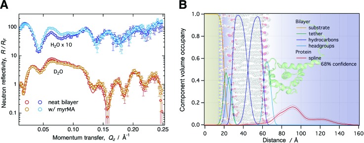
Fig. 1.
(a) NR curves normalized by the Fresnel reflectivity (RF) for a 50:50 DOPC:DOPS stBLM before and after protein addition (10 μM myrMA, pH 8.0, 50 mM NaCl). Each condition was characterized using two isotopically distinct bulk solvents (H2O and D2O-based buffer) using in situ buffer exchange. (b) CVO profiles for the bilayer structure and the membrane-associated protein were obtained by composition-space modeling. The protein orientation was determined by rigid body modeling using the NMR structure in PDB entry 2H3F. The background image visualizes the resulting protein orientation on the stBLM surface.

