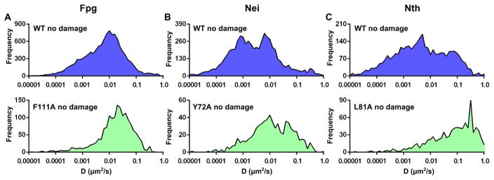
Fig. 5.
Diffusive behavior of WT (blue) and wedge variant (green) DNA glycosylases on undamaged lambda DNA. Columns A, B, and C show the time-weighted distribution of diffusive behavior for Fpg (A), Nei (B), and Nth (C). Details for these experiments can be found in Ref. [70].

