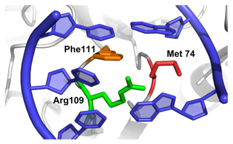
Fig. 6.

Crystal structure of the DNA helix following eversion of the damaged base into the active site of Fpg. The wedge residue (Phe111) is orange, while the other two residues in the intercalation loop Arg109 and Met 74 are green and red, respectively. (PDB 1K82).
