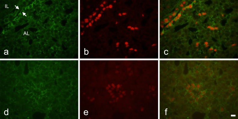
Fig. 2.
Double immunohistochemistry for Notch2 and SOX2 in adult rat pituitary gland. a–c) Frontal images around the marginal cell layers. d–f) Images of the main part of the anterior lobe. a and d) Notch2 immunoreactivity. b and e) SOX2 immunoreactivity. c and f) Merged images of a and b, and d and e, respectively. AL: anterior lobe, IL: intermediate lobe. Arrows: marginal cell layer. Bar = 10 μm.

