. 2017 Apr 30;5(1):24–29. doi: 10.14791/btrt.2017.5.1.24

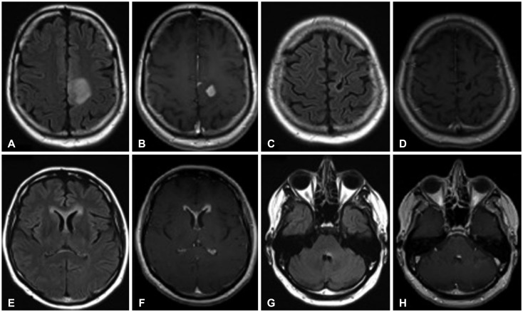
Fig. 3. 50-year-old male with WHO grade III astrocytoma (patient #10). Initial MRI examination demonstrates a FLAIR hyperintense (A) contrast-enhancing (B) lesion in the parasagittal posterior left frontal lobe. Follow-up examination after two years reveals no recurrent tumor at the resection site on FLAIR (C) or T1-weighted post-gadolinium images (D). However, at 26 months, there are new areas of subependymal FLAIR hyperintensity (E) and enhancement (F) in the lateral ventricles. Furthermore, there is a discrete focus of abnormal FLAIR hyperintensity (G) and enhancement (H) involving the posterior left pons, abutting the fourth ventricle. WHO, World Health Organization; MRI, magnetic resonance imaging; FLAIR, fluid attenuation inversion recovery.

Fig. 3