Figure 2.

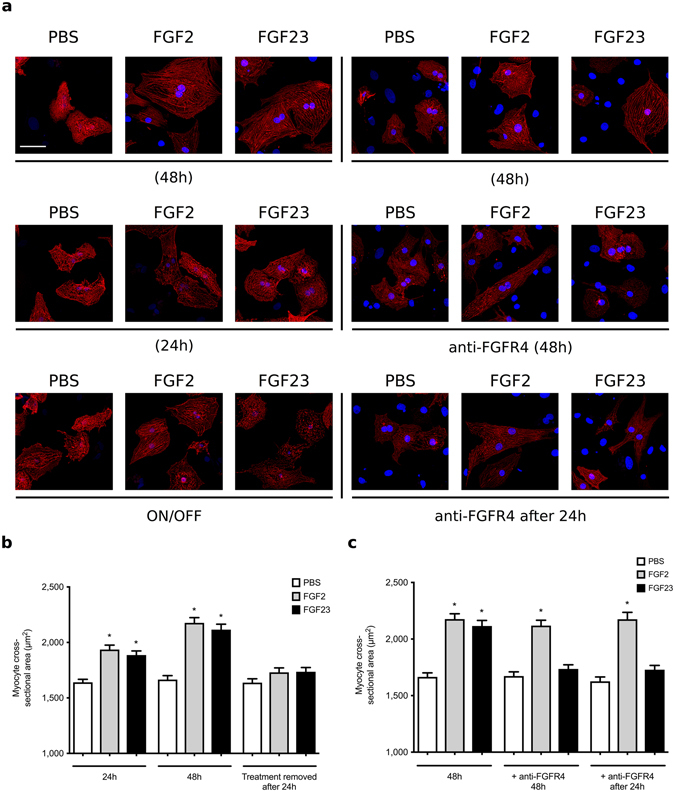
FGF23-induced hypertrophic growth of cultured cardiac myocytes is reversible. Analysis of hypertrophic cell growth in isolated neonatal rat ventricular myocytes (NRVM). Cells were treated with recombinant FGF23 or FGF2 at 25 ng/ml or with solvent (PBS) for various time periods. A subset of cells was co-treated with anti-FGFR4 at 10 µg/ml. Cross-sectional cell area was determined by confocal fluorescence microscopy based on α-actinin-positive immunostaining. (a) Representative immunofluorescence confocal images of isolated NRVMs. Myocytes are labeled with anti-α-actinin (red), and DAPI (blue) identifies nuclei (original magnification x63, scale bar 50 µm). (b) Within 24 and 48 hours of treatment, FGF23 and FGF2 significantly increase myocyte area. When NRVM are treated with FGFs for 24 hours, followed by FGF removal for 24 hours, cell area reverse back to levels of PBS-treated control cells. (c) When initiated at the same time, co-treatment with anti-FGFR4 blocks the development of FGF23-induced hypertrophy as analyzed after 48 hours (middle panel). When NRVM are treated with FGF23 for 24 hours and then co-treated with FGF23 and anti-FGFR4 for an additional 24 hours, myocyte area reverses back to control levels (right panel). Anti-FGFR4 co-treatment does not affect FGF2-induced hypertrophy. (150 cells per condition; n = 3 independent isolations of NRVM; *p < 0.05 compared with PBS).
