Figure 3.

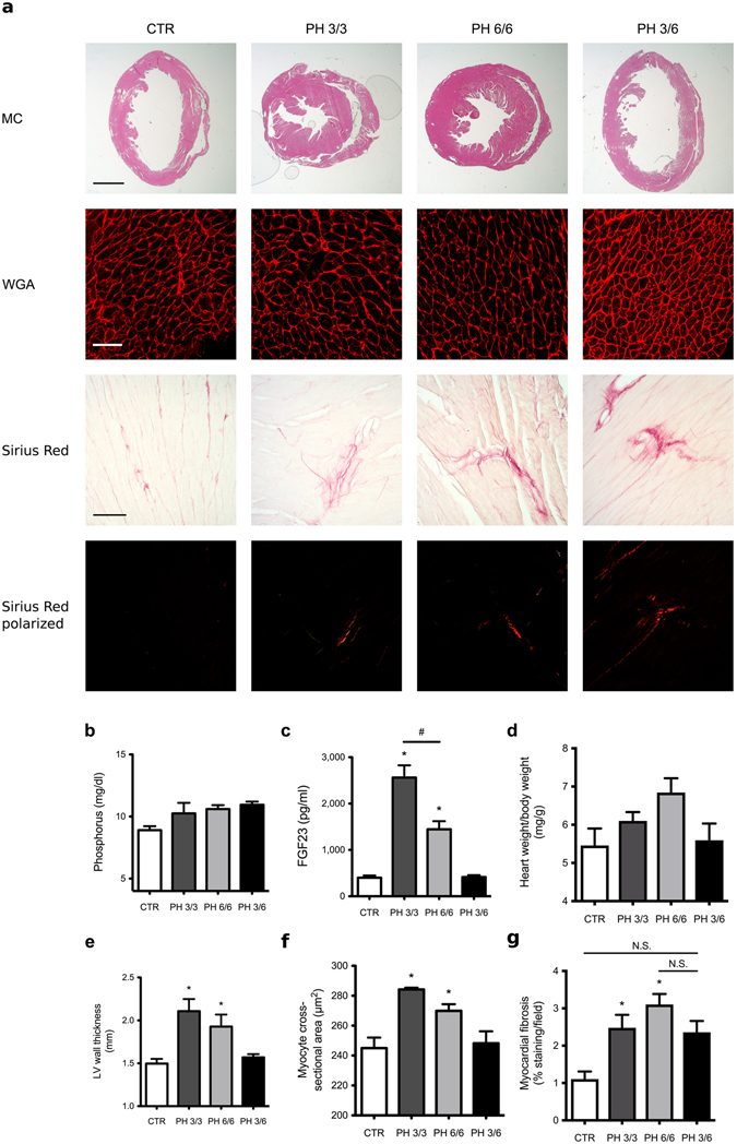
LVH induced by high phosphate diet is reversible in mice. Compared to normal chow (CTR), mice on a high phosphate diet for 3 months (PH 3/3) and 6 months (PH 6/6) develop LVH. Mice on a high phosphate diet for three months followed by a normal chow diet for three months (PH 3/6) do not develop LVH. (a) Upper panel: representative gross pathology of mid-chamber (MC) sections of the heart (H&E stain, original magnification x5, scale bar 2 mm). Middle panel: representative WGA-stainings of the LV free wall (original magnification x63, scale bar 50 µm). Lower two panels: representative Picrosirius Red stainings demonstrating fibrosis in the LV free wall (original magnification x10, scale bar 100 µm). (b) Compared to CTR, mice on a high phosphate diet (PH 3/3, PH 6/6 and PH 3/6) show a trend towards higher serum phosphate levels. (c) Serum FGF23 levels are significantly increased in animals fed a high phosphate diet (PH 3/3, PH 6/6) and return to normal levels when mice are transitioned to normal chow for three months (PH 3/6). (d) Mice on high phosphate diet exhibit a trend towards increased ratios of heart weight to body weight. (e) Average LV wall thickness as measured by seven individual angles at 0, 30, 60, 90, 120, 150, 180 degrees along the LV free wall from the hemi-circle of the short axis. (f) Cross-sectional area of 25 myocytes per field of 4 fields along the LV free wall. (g) Quantification of myocardial fibrosis (8 fields of view around the LV). All values are mean ± SEM (n = 4–9 per group; *p < 0.05 compared with CTR, #p < 0.01 between groups).
