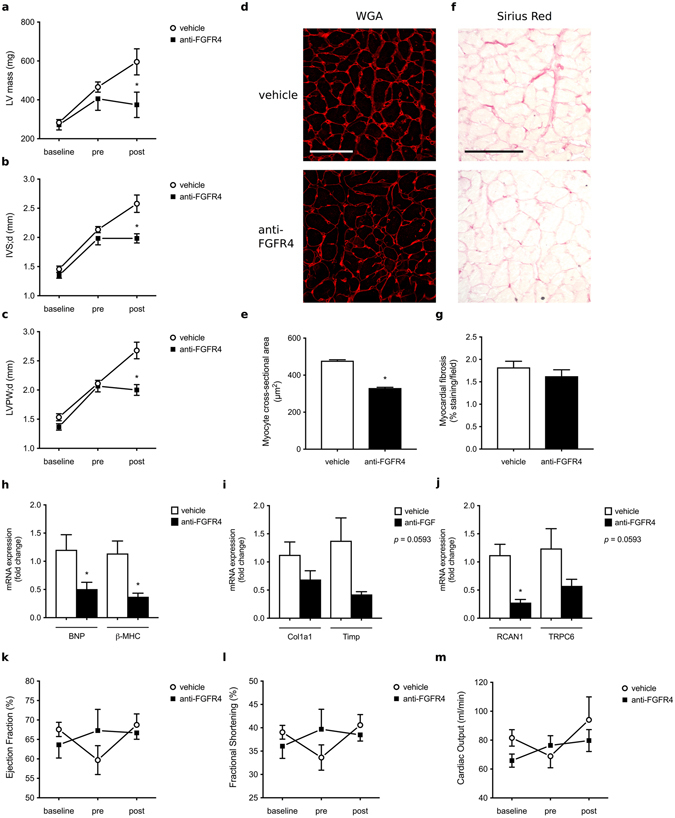
Figure 4.

FGFR4 blockade inhibits the progression of LVH in the 5/6 nephrectomy rat model of CKD. (a–c) Cardiac parameters of 5/6 nephrectomized rats were evaluated by echocardiography at baseline, four weeks (pre) and eight weeks after surgery (post). All 5/6 nephrectomized animals develop LVH four weeks post-surgery, but anti-FGFR4 treatment prevents the progression of cardiac remodeling when compared to vehicle-treated rats. (d) Representative images of WGA-stainings of the LV free wall (original magnification x63, scale bar 70 µm). (e) Cross-sectional area of 25 myocytes per field of 4 fields along the LV free wall. (f) Representative Picrosirius Red stainings of the LV free wall (original magnification x20, scale bar 100 µm). (g) Quantification of myocardial fibrosis based on positive Picrosirius Red labeling (8 fields of view around the LV). (h,i,j) Quantitative real-time PCR analysis of cardiac tissue using GAPDH as a house keeping gene. mRNA levels of hypertrophic markers (BNP, β-MHC) and the NFAT target gene RCAN1 are significantly decreased in anti-FGFR4 treated rats when compared to vehicle-injected animals. Fibrosis markers (Col1a1, Timp) and the NFAT target gene TRPC6 show a trend towards a decrease upon FGFR4 blockade (p = 0.0593). (k,l,m) Cardiac function remains unchanged upon induction of CKD as well as before and after administration of anti-FGFR4. No significant differences were found in ejection fraction, fractional shortening and cardiac output over time. All values are mean ± SEM (n = 6–8 per group; *p < 0.05).
