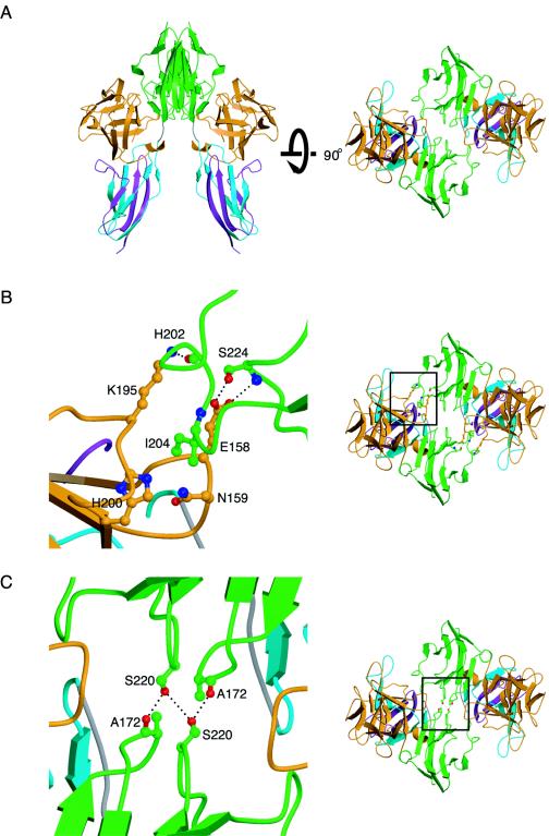
FIG. 2.
Crystallographic FGF10-FGFR2b dimer interface. (A) Ribbon diagram of the twofold crystallographic FGF10-FGFR2b canyon dimer in two views related by a 90° rotation about the horizontal axis. (B) Secondary interaction site at the dimeric interface. (C) Interactions at the receptor-receptor interface. D2, D3, and FGF are colored as in Fig. 1. The side chains of selected interacting residues are displayed. Oxygen atoms are colored red, nitrogen is blue, and carbon atoms are the same color as the molecules to which they belong. On the right are views of the whole structure in the exact orientation that the detailed views show, with the regions of interest boxed.

