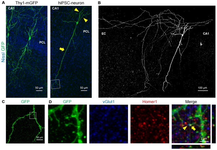
Figure 4.
hiPSC-neurons transplanted in the PCL exhibited pyramidal cell-like dendrites with synaptic spines. (A) Representative images of a Thy1-mGFP+ pyramidal cell in the CA1 PCL at 11 DIV (left) and a GFP+ hiPSC-neuron in the PCL at 7 DPT (right). (B) A whole image of the GFP+ hiPSC-neuron in (A). The GFP+ hiPSC-neuron projected its axons to the EC. (C) A magnified image of the boxed square in (A, right) revealed multiple spines along the dendrite. (D) Representative immunohistochemical images of the boxed square in (C). The spine of the GFP+ hiPSC-neuron (arrows) was colocalized with the postsynaptic marker Homer1 and the presynaptic marker vGlut1 (arrowheads).

